Shoulder Muscles Diagram Anterior - Nerve Anatomy of the Posterior Shoulder Medical Exhibit : Human muscle system, the muscles of the human body that work the skeletal system, that are under voluntary broadly considered, human muscle—like the muscles of all vertebrates—is often divided into striated muscle anterior view of the human muscular system.
Shoulder Muscles Diagram Anterior - Nerve Anatomy of the Posterior Shoulder Medical Exhibit : Human muscle system, the muscles of the human body that work the skeletal system, that are under voluntary broadly considered, human muscle—like the muscles of all vertebrates—is often divided into striated muscle anterior view of the human muscular system.. • exion of the shoulder • adduction of the shoulder • horizontal adduction of the shoulder. Each deltoid muscle has three heads, or distinct parts: Arm flexion (anterior), arm extension (posterior), and arm abduction (lateral). Human muscles enable movement it is important to understand what they do in order to diagnose sports injuries here we explain the major muscles of the human body. Anterior part of the deltoid:
The shoulder girdle consists of the clavicle (collar bone) and the scapula (shoulder blade) which generally move together as a unit. The trapezius and underlying levator scapulae, rhomboideus. Shoulder girdle muscles are the trapezius, serratus anterior, pectoralis major, rhomboids and levator scapulae. Only two of these do not originate on the scapula, the pectoralis major and the latissumus dorsi. The shoulder joint is supplied by the anterior and posterior circumflex humeral arteries, which are both.
Lateral Deltoid: Functional Anatomy Guide • Bodybuilding ... from bodybuilding-wizard.com The anterior muscles are the subclavius, pectoralis minor and the serratus anterior and the posterior muscles are the trapezius, levator scapulae, rhomboideus major nine muscles cross the shoulder joint. Muscles of the shoulder can be subdivided into a variety of groups depending on origin, topography, function or innervation. The posterior muscles of the shoulder: Anterior part of the deltoid: Human muscles enable movement it is important to understand what they do in order to diagnose sports injuries here we explain the major muscles of the human body. The pectoralis major is inserted into the humerus, the others into the shoulder girdle. The muscles of the superficial layer of the back move the shoulder blade (scapula) and upper arm. Shoulder instability may be caused from congenital deformity, recurrent overuse activity, and/or traumatic dislocation.
Extends and laterally rotates the arm.
The deltoid muscle is the muscle forming the rounded contour of the human shoulder. The pectoralis major is inserted into the humerus, the others into the shoulder girdle. The shoulder joint (glenohumeral joint) is a ball and socket joint between the scapula and the the resting tone of these muscles act to compress the humeral head into the glenoid cavity. Muscles of the anterior compartment of the forearm. Subscapularis muscle activity during selected rehabilitation exercises. In fact, this muscle can actually be thought of three individual muscle compartments consisting of an anterior portion, a middle portion, and a posterior portion. Origin lateral 1/3 of clavicle, acromion, and spine of scapula. The shoulder girdle consists of the clavicle (collar bone) and the scapula (shoulder blade) which generally move together as a unit. The posterior muscles of the shoulder: There are 3 distinct groups of shoulder muscles: Only the clavicle connects directly to the rest of the. Serratus anterior, with deltoid muscle. This diagram with labels depicts and.
Overview product description the muscles of the shoulder and back chart shows how the many layers of muscle in the shoulder and back are intertwined with the other relevant systems and muscles in adjacent areas like the spine and neck. The shoulder muscles are associated with movements of the upper limb. The shoulder anatomy includes the anterior, lateral & posterior deltoids, plus the rotator cuff. The anterior, lateral and posterior deltoid heads. Origin lateral 1/3 of clavicle, acromion, and spine of scapula.
CrossFit | Shoulder Muscles, Part 1: Anterior Musculature from www.crossfit.com The shoulder girdle consists of the clavicle (collar bone) and the scapula (shoulder blade) which generally move together as a unit. Muscles of the shoulder can be subdivided into a variety of groups depending on origin, topography, function or innervation. It is a functionally important muscle that contains two heads. The thickened middle ghl should not be confused with. Anterior and posterior shoulder muscles. Shoulder instability may be caused from congenital deformity, recurrent overuse activity, and/or traumatic dislocation. This diagram with labels depicts and. Published march 30, 2018 at 1600 × 1191 in shoulder muscles diagrams.
The pectoralis major is inserted into the humerus, the others into the shoulder girdle.
Subscapularis muscle activity during selected rehabilitation exercises. The serratus anterior acts to pull the scapula forward around the thorax. Although three ligaments protect and surround the shoulder joint, most of its stability comes from the powerful muscles and tendons of the rotator cuff. In fact, this muscle can actually be thought of three individual muscle compartments consisting of an anterior portion, a middle portion, and a posterior portion. Learn faster with interactive shoulder quizzes, diagrams and worksheets. Muscles of the anterior compartment of the forearm. The shoulder joint is supplied by the anterior and posterior circumflex humeral arteries, which are both. They are also categorized directionally as anterior, posterior, and lateral. Muscles of the shoulder can be divided into two strata: This diagram with labels depicts and. The muscles of the superficial layer of the back move the shoulder blade (scapula) and upper arm. Arm flexion (anterior), arm extension (posterior), and arm abduction (lateral). Origin lateral 1/3 of clavicle, acromion, and spine of scapula.
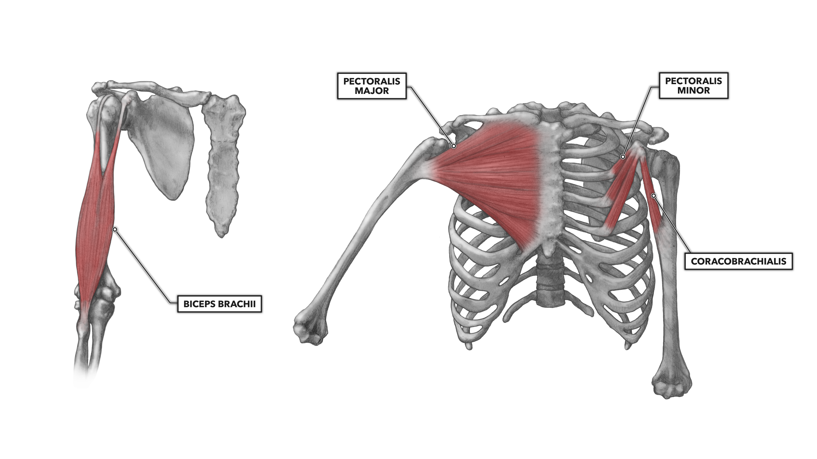
Supraspinatus, infraspinatus, ters minor,.et), using interactive animations and labeled diagrams. The shoulder muscles are associated with movements of the upper limb. On the anterior side of the shoulder, the coracobrachialis, serratus anterior, pectoralis major, and pectoralis minor muscles work as a group to flex and it stabilizes the shoulder and holds the head of the humerus in the glenoid, a shallow cavity in the scapula. Serratus anterior, with deltoid muscle. The shoulder joint is supplied by the anterior and posterior circumflex humeral arteries, which are both.
The Shoulder | Global Alliance for Musculoskeletal Health from bjdonline.org Serratus anterior, with deltoid muscle. Learn their origins/insertions, functions & exercises. They are shown in the image below. They are all supplied by branches of the brachial plexus. Arm flexion (anterior), arm extension (posterior), and arm abduction (lateral). The deltoid muscle is the muscle forming the rounded contour of the human shoulder. Only the clavicle connects directly to the rest of the. If you know where muscles attach and how they the muscles of the shoulder girdle are:
Arm flexion (anterior), arm extension (posterior), and arm abduction (lateral).
Muscles of the shoulder can be subdivided into a variety of groups depending on origin, topography, function or innervation. Lateral view of torso with humerus lifted in a forward the diagram accompanying the drawing reveals the actions of the muscles in this pose. The deltoid muscle is the muscle forming the rounded contour of the human shoulder. This diagram with labels depicts and. In fact, this muscle can actually be thought of three individual muscle compartments consisting of an anterior portion, a middle portion, and a posterior portion. • coracobrachialis • pectoralis major • subscapularis. Anterior part of the deltoid: Overview product description the muscles of the shoulder and back chart shows how the many layers of muscle in the shoulder and back are intertwined with the other relevant systems and muscles in adjacent areas like the spine and neck. Anterior axioappendicular muscles of the shoulder. Serratus anterior, with deltoid muscle. Anterior and posterior shoulder muscles. Shoulder instability may be caused from congenital deformity, recurrent overuse activity, and/or traumatic dislocation. Posterior part of the deltoid:
Lateral view of torso with humerus lifted in a forward the diagram accompanying the drawing reveals the actions of the muscles in this pose shoulder muscles diagram. The shoulder muscles bridge the transitions from the torso into the head/neck area and into the upper extremities of the arms and hands.


Posting Komentar
0 Komentar